Skip Navigation
Skip to contents

JSIE : Journal of Surgical Innovation and Education

OPEN ACCESS
SEARCH
Search

Articles

Page Path
HOME > J Surg Innov Educ > Volume 2(2); 2025 > Article
How I Do It
Laparoscopic Inguinal Hernia Repair in Female Pediatric Patients
Joong Kee Youn1,2orcid
Journal of Surgical Innovation and Education 2025;2(2):35-38.
DOI: https://doi.org/10.69474/jsie.2025.00325
Published online: December 19, 2025

1Department of Pediatric Surgery, Seoul National University Hospital, Seoul, Republic of Korea

2Department of Surgery, Seoul National University College of Medicine, Seoul, Republic of Korea

Corresponding author: Joong Kee Youn, MD, PhD Department of Pediatric Surgery, Seoul National University Hospital, 101 Daehak-ro, Jongno-gu, Seoul 03080, Republic of Korea Tel: +82-2-2072-4252, Fax: +82-2-747-5130, E-mail: bead47@snu.ac.kr
• Received: November 11, 2025   • Revised: December 10, 2025   • Accepted: December 10, 2025

© 2025 Korean Surgical Skill Study Group

This is an Open Access article distributed under the terms of the Creative Commons Attribution Non-Commercial License (http://creativecommons.org/licenses/by-nc/4.0) which permits unrestricted non-commercial use, distribution, and reproduction in any medium, provided the original work is properly cited.

prev next
  • 0 Views
  • 2 Download
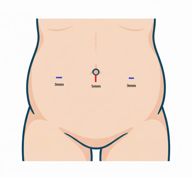
  • Laparoscopic inguinal hernia repair (LIHR) has become a widely adopted surgical approach for pediatric inguinal hernia because it offers superior visualization and enables simultaneous assessment of the contralateral side. An 8-month-old female infant presented with a spontaneously reducing bulge in the right inguinal region, most noticeable during diaper changes. Physical examination revealed an easily reducible inguinal mass that was palpably firm and spherical, raising strong suspicion for ovarian inclusion. A positive silk glove sign was also identified on the contralateral side. Under general anesthesia, a three-port laparoscopic technique was used, consisting of a 5-mm umbilical port and two 3-mm working ports. The peritoneum and gubernaculum were carefully dissected from surrounding structures using electrocautery before sac closure, a step performed to minimize the risk of recurrence. Both the symptomatic right hernia sac and the asymptomatic contralateral patent processus vaginalis, which was visually confirmed intraoperatively, were closed using an intracorporeal purse-string high ligation with absorbable sutures. Three-port LIHR with meticulous dissection of the peritoneum and gubernaculum represents an effective and definitive technique for pediatric inguinal hernia repair. This approach allows simultaneous bilateral repair and is associated with excellent postoperative recovery, supporting its continued use as a primary surgical method.
The cumulative incidence of inguinal hernia from birth to 15 years of age has been reported as 6.62% in boys and 0.74% in girls in a nationwide cohort study with incarceration rate around 1%–2% [1]. While traditional open herniorrhaphy remains an established approach, laparoscopic techniques have increasingly gained acceptance since the pioneering work by Esposito and Montupet [2] in the mid-1990s.
Laparoscopic inguinal hernia repair (LIHR) is now widely applied and has become a major surgical approach due to several reported advantages over open surgery, including superior visualization of the inguinal anatomy, minimal invasiveness and excellent cosmesis, and the ability to assess and simultaneously repair a contralateral patent processus vaginalis (PPV) [3].
This report details the three-port technique, which currently remains one of the most commonly practiced methods for this procedure.
Patient
An 8-month-old female infant presented to our outpatient clinic. One week prior to the visit, the patient’s parents had noticed a bulging, protruding mass in the right inguinal region. They reported that the mass protruded when the infant cried but subsequently reduced spontaneously. The patient was initially taken to a local pediatric clinic where an inguinal hernia was suspected, leading to a referral to our pediatric surgery department.
On physical examination, a bulging, protruding mass was clearly visible in the right inguinal region. When the patient was laid supine on the examination bed, the mass was easily reducible. Palpation of the contralateral side revealed no evidence of herniation; however, a positive silk glove sign was noted. The patient was scheduled for an elective LIHR and was subsequently discharged home.
Surgical procedure
The procedure was initiated under general anesthesia. An open technique was used to insert a 5-mm infra-umbilical port for the introduction of a 5-mm flexible scope. Subsequently, two working ports were placed: two 3-mm trocars were inserted at the right and left midclavicular lines lateral to the umbilicus (Fig. 1). In infants and small babies, the working ports were positioned more caudally than the umbilical level to facilitate optimal instrument maneuverability.
Upon insertion of the laparoscopic camera, the asymptomatic contralateral internal inguinal ring (IIR) was routinely inspected, which revealed a PPV. If ovary was herniated, immediate reduction should be performed before mobilization of the sac. Prior to defect closure, the peritoneum surrounding the IIR was circumferentially incised and meticulously dissected from underlying structures using electrocautery to ensure complete mobilization of the hernia sac neck, including the gubernaculum, without bleeding to minimize the risk of recurrence. This was initiated by gently elevating the peritoneum and making a small entry point. The opening was then gradually enlarged carefully mobilizing under direct visualization. In open pediatric inguinal hernia repair, it has long been standard practice to ligate and divide the hernia sac together with the round ligament at the level of the internal ring. When this principle is applied to laparoscopic hernia repair, the same step can be safely reproduced intraperitoneally by performing a meticulous transection of the gubernaculum. The definitive repair consisted of a laparoscopic high ligation achieved by placing a purse-string suture at the deepest margin of the peritoneal defect. Absorbable 5/0 sutures (Vicryl®) were used, and the knot was secured via intracorporeal ligation. The contralateral PPV was also ligated in the same manner (Video). To prevent a future metachronous contralateral hernia, it is considered reasonable to close a detected contralateral PPV during the same anesthesia.
Following successful closure of the internal rings, the pneumoperitoneum was released by deflating the abdomen, and all abdominal incisions were closed in layers.
Postoperative management
In line with the standard protocol, the patient was discharged on the same day of the operation. Follow-up examinations were scheduled in the outpatient clinic at 2 weeks and 3 months postoperatively.
Ethical approval
This study was reviewed and approved by the Institutional Review Board (IRB) of Seoul National University Hospital (IRB No. 2212-082-1386). The requirement for informed consent was waived because of the retrospective nature of the study, the use of anonymized data, and the absence of any additional risk to the participants.
Many surgical techniques for LIHR in pediatric patients have been described in the literature. These techniques can generally be summarized as intracorporeal, extracorporeal, and percutaneous approaches [4].
For instance, a single-port percutaneous method has been reported in various countries, including Japan and European nations. This technique involves placing only one port below the umbilicus solely for the camera. A small skin incision is then made in the inguinal area, allowing a suture needle to be passed from outside the abdomen to perform a simple suture closure of the IIR. This method is widely employed due to its significant advantages, such as excellent cosmetic outcomes and shorter operative time [5].
Currently, LIHR is being applied far more frequently than the traditional open method and is widely considered the main surgical approach for pediatric inguinal hernia. In the early stages of its adoption, several issues were raised against LIHR compared to open surgery, including longer operative time and a higher recurrence rate [6]. These concerns, however, have been gradually resolved. While initial reports showed a significant difference in operative time (e.g., 25 min versus 47 min), the literature indicated a gradual decrease with advancements in laparoscopic training [7]. Furthermore, in LIHR, the ovary and uterus were frequently observed to be shifted toward the hernia orifice, which highlights a potential benefit of the laparoscopic approach in reducing the risk of injury to the ovary, fallopian tube, and potentially the uterus [8].
Several modifications of the technique have been described and published, including the Z suture, and the use of interrupted or continuous sutures. However, these techniques were not without a high recurrence rate due to the “skip area” left over the vas deferens and testicular vessels [6]. Consequently, many techniques were introduced in which the peritoneum was transected or disconnected at the internal ring, aiming to create some trauma and scarring to improve healing [9]. The recurrence rate was successfully decreased to 0%–4% in many series using these techniques [10].
Fig. 1.
The size and position of laparoscopic ports applied in pediatric laparoscopic inguinal hernia repair.
jsie-2025-00325f1.jpg
Video.
­
  • 1. Chang SJ, Chen JY, Hsu CK, Chuang FC, Yang SS. The incidence of inguinal hernia and associated risk factors of incarceration in pediatric inguinal hernia: a nation-wide longitudinal population-based study. Hernia. 2016;20:559-563.ArticlePubMedPDF
  • 2. Esposito C, Montupet P. Laparoscopic treatment of recurrent inguinal hernia in children. Pediatr Surg Int. 1998;14:182-184.ArticlePubMed
  • 3. Gorsler CM, Schier F. Laparoscopic herniorrhaphy in children. Surg Endosc. 2003;17:571-573.ArticlePubMedPDF
  • 4. Ostlie DJ, Ponsky TA. Technical options of the laparoscopic pediatric inguinal hernia repair. J Laparoendosc Adv Surg Tech A. 2014;24:194-198.ArticlePubMed
  • 5. Oue T, Kubota A, Okuyama H, Kawahara H. Laparoscopic percutaneous extraperitoneal closure (LPEC) method for the exploration and treatment of inguinal hernia in girls. Pediatr Surg Int. 2005;21:964-968.ArticlePubMedPDF
  • 6. Shalaby R, Ismail M, Dorgham A, Hefny K, Alsaied G, Gabr K, et al. Laparoscopic hernia repair in infancy and childhood: evaluation of 2 different techniques. J Pediatr Surg. 2010;45:2210-2216.ArticlePubMed
  • 7. Esposito C, Escolino M, Cortese G, Aprea G, Turrà F, Farina A, et al. Twenty-year experience with laparoscopic inguinal hernia repair in infants and children: considerations and results on 1833 hernia repairs. Surg Endosc. 2017;31:1461-1468.ArticlePubMedPDF
  • 8. Muta Y, Odaka A, Inoue S, Takeuchi Y, Beck Y. Female pediatric inguinal hernia: uterine deviation toward the hernia side. Pediatr Surg Int. 2021;37:1569-1574.ArticlePubMedPDF
  • 9. Oshiba A, Ashour K, Aboheba M, Shehata S, Shalaby R. Comparative study between purse–string suture and peritoneal disconnection with ligation techniques in the laparoscopic repair of inguinal hernia in infants and children. Ann Pediatr Surg. 2016;12:137-141.Article
  • 10. Boo YJ, Han HJ, Ji WB, Lee JS. Laparoscopic hernia sac transection and intracorporeal ligation show very low recurrence rate in pediatric inguinal hernia. J Laparoendosc Adv Surg Tech A. 2012;22:720-723.ArticlePubMed

Figure & Data

References

    Citations

    Citations to this article as recorded by  

      • PubReader PubReader
      • ePub LinkePub Link
      • Cite this Article
        Cite this Article
        export Copy Download
        Close
        Download Citation
        Download a citation file in RIS format that can be imported by all major citation management software, including EndNote, ProCite, RefWorks, and Reference Manager.

        Format:
        • RIS — For EndNote, ProCite, RefWorks, and most other reference management software
        • BibTeX — For JabRef, BibDesk, and other BibTeX-specific software
        Include:
        • Citation for the content below
        Laparoscopic Inguinal Hernia Repair in Female Pediatric Patients
        J Surg Innov Educ. 2025;2(2):35-38.   Published online December 19, 2025
        Close
      • XML DownloadXML Download
      Figure
      • 0
      Laparoscopic Inguinal Hernia Repair in Female Pediatric Patients
      Image
      Fig. 1. The size and position of laparoscopic ports applied in pediatric laparoscopic inguinal hernia repair.
      Laparoscopic Inguinal Hernia Repair in Female Pediatric Patients

      JSIE : Journal of Surgical Innovation and Education
      TOP